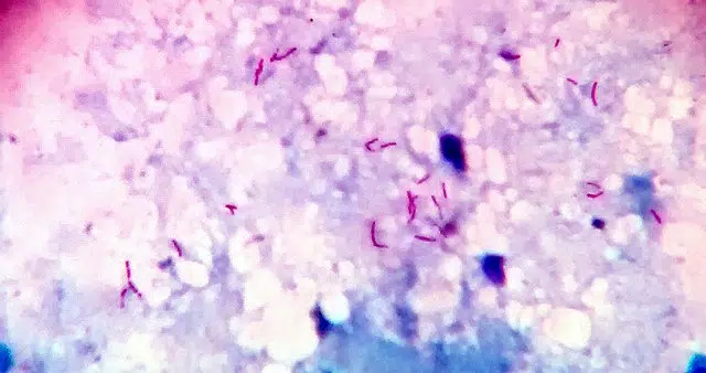
Smear microscopy is a laboratory procedure used to analyze cells and microorganisms present in a biological sample. Typically, a smear is made from a sample of blood, urine, vaginal secretion, or other material, which is smeared onto a glass slide and stained for easy viewing under a microscope.
This method is widely used in the healthcare field to diagnose various diseases, such as bacterial, fungal and viral infections, as well as to assist in monitoring treatments and monitoring health conditions.
There are different types of smear microscopy, such as blood smear microscopy, which identifies blood cells and possible abnormalities, and vaginal smear microscopy, used to detect gynecological infections, for example. Each type of smear requires specific sample preparation and a suitable staining technique for microscopic visualization.
Step by step guide to performing the smear correctly and effectively.
To perform a smear correctly and effectively, it's important to follow a few simple steps. A smear is a technique used in microscopy to prepare cell or tissue samples for microscopic examination. See the step-by-step process for performing a smear below:
Step 1: First, clean the glass blade with 70% alcohol and let it dry completely. Make sure the blade is free of dust or any residue.
Step 2: Next, take the sample you wish to analyze and place a small amount on the glass slide. Spread the sample evenly, creating a thin layer.
Step 3: With the help of another glass slide, make the smear. Place the slide at an angle of approximately 45 degrees and slide the slide over the sample with smooth, even movements.
Step 4: Allow the smear to air dry for a few minutes. Avoid blowing on or heating the slide, as this may alter the sample.
Step 5: Finally, fix the smear with methyl alcohol or another fixative. Allow it to dry completely before observing it under a microscope.
By following these simple steps, you'll be able to perform the smear correctly and obtain accurate results in the microscopic analysis of the sample. Remember the importance of proper technique to ensure the quality of the results obtained.
What should the ideal smear look like?
Smear microscopy is a widely used procedure in healthcare to analyze cells present in different types of biological samples. For the analysis to be accurate, it is essential that the smear has an ideal appearance, with specific characteristics that facilitate the visualization and identification of the cells present.
An ideal smear should have well-distributed and evenly spaced cells across the slide. The cells should be intact, with no signs of degeneration or rupture. Furthermore, the smear's color should be homogeneous, allowing for better visualization of cellular structures.
The cells present in the smear must be well preserved and maintain their morphological characteristics. This means that the cells must maintain their original shape and not present significant alterations that could hinder correct identification.
It's important to emphasize that the quality of the smear is essential for an accurate and reliable analysis. Therefore, it's crucial to follow the smear preparation procedure correctly and ensure all steps are performed correctly.
By following these guidelines, it is possible to obtain more accurate and reliable results in the microscopic analysis of biological samples.
How important is the smear?
The smear is a fundamental procedure in microscopy. It involves preparing a sample for analysis under a microscope, allowing for detailed visualization of cells, microorganisms, and other structures. The importance of the smear lies precisely in enabling the observation of elements that would not be visible to the naked eye.
To perform a smear, the sample must be spread evenly and gently on a glass slide, ensuring that the cells are evenly distributed. The material is then fixed and stained to facilitate identification of structures under a microscope. There are different types of smears, such as blood smears, secretion smears, and culture smears.
Smear microscopy can be used to diagnose various diseases, identify infectious agents, assess blood cell quality, and monitor treatments, among other applications. Therefore, smear microscopy is a crucial step in performing laboratory tests and obtaining important information about a patient's health.
Without the smear, much valuable information could not be obtained, compromising the accuracy and effectiveness of laboratory tests.
What is a fine smear for and how important is it for women's health?
Smear microscopy is an important procedure in women's health, essential for diagnosing various diseases. One of the most well-known tests is the thin smear, which involves collecting cells from the cervix for laboratory analysis.
A thin smear is primarily used to detect changes in cervical cells, such as precancerous and cancerous lesions. These changes can be caused by the Human Papillomavirus (HPV), one of the main risk factors for the development of cervical cancer.
It is important to emphasize that the thin smear is a preventive examination that allows for the identification of cellular changes in their early stages, enabling more effective treatment and increasing the chances of a cure. Furthermore, the examination is also fundamental for monitoring women who have already been diagnosed with cervical lesions.
Therefore, it is essential that women regularly undergo a thin smear test , as recommended by their doctor. The test is simple, painless, and can save lives, being a fundamental tool in the prevention of cervical cancer.
Smear microscopy: procedure, purpose and types
A blood smear is a diagnostic tool for active pulmonary tuberculosis. It is a laboratory technique that allows the detection and measurement of acid-fast bacilli, such as Mycobacterium tuberculosis . It is the most widely used method in the world for diagnosing active pulmonary tuberculosis in the community and evaluating the benefits of treatment.
Pulmonary tuberculosis is a contagious disease caused by Mycobacterium tuberculosis that infects the lungs, but can affect other organs and systems. Globally, more than 6 million people suffer from pulmonary tuberculosis (TB).
Mycobacterium tuberculosis and other non-tuberculous Mycobacterium species have a lipid coating on their cell wall rich in mycolic acid, which is resistant to discoloration with acid alcohol after staining with basic dyes.
Therefore, special staining methods, such as the Ziehl-Neelsen method, are used. These simple and inexpensive methods allow the cell wall to retain the stain by washing the sample with an acidic solution, and the Mycobacterium cell wall acquires a red coloration as opposed to the blue color of other bacteria.
A swab sample requires a sputum sample, preferably collected in the morning upon waking. Tissue or body fluid samples can also be used.
Production
The samples to be processed for smear microscopy are sputum samples, tissue samples, or body fluid samples. Tissue samples can be removed by bronchoscopy or by biopsy of lymph nodes or other tissues.
As tuberculosis can affect any organ or system, samples can be very varied, among which can be examined: urine samples, cerebrospinal fluid, pleural fluid, ascitic fluid, blood, pus from open cavities, biopsies, etc.
Samples from extrapulmonary lesions should also be processed for culture. Sputum samples are collected in the morning after ascent.
Multiple samples can be collected consecutively or on consecutive days. Samples should be labeled in closed, wide-mouthed containers. They should include patient information, sample type, and collection date.
Technical procedure
Once the sample is obtained, it can be centrifuged to concentrate or used directly. The initial procedure is to place a few drops of the sample and spread them on a slide.
– The sample is covered with filtered fuchsin (dye solution).
– Then it is heated until it produces about three whitish vapor emissions.
– Wait five minutes for the sample to cool and wash with water.
– Cover with bleaching solution (acidic alcohol) for a period of 2 minutes.
– It is washed again with water.
– The sample is covered with methylene blue and left to stand for one minute.
– It is washed with water.
– Let it air dry and proceed to observation under the microscope.
This procedure is quick, simple, inexpensive, and allows Mycobacterium tuberculosis cells to stain red . This technique is called the Ziehl-Neelsen staining technique.
Microscopic observation consists of identifying the presence of acid-fast bacilli and their quantification by field.
Mycobacterium tuberculosis cells appear as red, granular, curved rods on a blue background. They may be isolated, paired , or clustered. Observation should be done by field, and at least 100 fields should be examined for each smear.
The result is considered positive when there are more than 10 cells of M. tuberculosis in each field . This corresponds to 5000 to 100,00 bacilli per milliliter of sample.
In these cases, it is considered positive for Mycobacterium tuberculosis , since non-tuberculous Mycobacterium or other acid-resistant bacteria have different shapes and the value observed per field is generally less than 10.
The person examining the sample must have experience or knowledge in detecting and recognizing these microorganisms, as there may be false negatives or positives in the results.
There are other fluoroscopic techniques that are more sensitive but much more expensive. One such technique uses Auramine-O staining to obtain green fluorescence, or Auramine O/Rhodamine B staining to observe yellow/orange fluorescence.
Positive cases are usually cultured and antibiogram performed to confirm the diagnosis and appropriate treatment selection.
What is bacilloscopy for?
Tuberculosis is an infectious disease that affects the lungs and other organs and organ systems of the body. It is a contagious disease that causes severe damage to the respiratory system that, if not properly treated, can lead to death.
Tuberculosis is transmitted almost exclusively through airborne sputum from a patient infected with pulmonary tuberculosis. These tiny droplets can linger in the air and infect the person who inhales them.
Infection requires prolonged contact with an infected patient who continually expectorates bacilli in a poorly ventilated environment. People with immunodeficiencies are more prone to infection.
HIV patients are more likely than the general population to develop serious respiratory infections, including pulmonary tuberculosis. Hence the importance of smear microscopy as a rapid and inexpensive diagnostic method for tuberculosis.
Bacilloscopy allows the bacteriological diagnosis of tuberculosis to be made, however, a positive result must be confirmed with other studies.
Studies that confirm the diagnosis of tuberculosis include radiological examinations that show characteristic images in the upper lobes of the lung, positive tuberculin tests, and positive sample cultures.
Negative smear microscopy
A negative smear does not necessarily rule out a diagnosis of tuberculosis, as bacilli are not removed consistently, especially when the sample is sputum. Therefore, in these cases, when the diagnosis is suspected, serial samples are required.
Positive bacilloscopy
A positive sputum smear microscopy is indicative of tuberculosis and should be confirmed with other complementary studies. Whenever the result is positive, a culture and antibiogram should be performed.
Once treatment is established, a sustained decrease in the smear positivity scale until negative results are obtained allows the benefits of treatment to be monitored.
References
- Aziz, MA (2002). External quality assessment for AFB microscopy.
- Desikan, P. (2013). Sputum microscopy in tuberculosis: is it still relevant? The Indian Journal of Medical Research , 137 (3), 442.
- Fauci, A.S., Kasper, D.L., Hauser, S.L., Jameson, J.L., & Loscalzo, J. (2012). Harrison's Principles of Internal Medicine (Vol. 2012). DL Longo (Ed.). New York: McGraw-Hill.
- Ngabonziza, J.C.S., Ssengooba, W., Mutua, F., Torrea, G., Dushime, A., Gasana, M., … and Muvunyi, C.M. (2016). Diagnostic performance of smear microscopy and incremental yield of Xpert in detecting pulmonary tuberculosis in Rwanda. BMC Infectious Diseases , 16 (1), 660.
- Sardiñas, M., García, G., Rosários, MM, Díaz, R. and Mederos, LM (2016). Importance of quality control of bacilloscopy in laboratories that perform tuberculosis diagnosis. Chilean Journal of Infectious Diseases: official journal of the Chilean Society of Infectious Diseases , 33
- Sequeira de Latini, MD, & Barrera, L. (2008). Manual for the bacteriological diagnosis of tuberculosis: standards and technical guide: part 1 bacilloscopy. No. Manual for the bacteriological diagnosis of tuberculosis: standards and technical guide: part 1 sputum smear microscopy (pp64-64).